Mosman Cardiology
  • Home
  • About
  • Cardiologist Team
    • Dr Amely Zaininger
    • Dr Elizabeth Robertson
    • Professor Mark McGuire
    • Dr Caroline Medi
    • Dr Kevin Liou
    • Dr Kevan Karimi
    • Dr Lucy McGrath-Cadell
    • Dr Katy Ellenberger
    • Associate Professor Rebecca Kozor
  • Facilities and Services
  • For Patients
    • Stress testing
    • Echocardiogram
    • Exercise Stress Echocardiogram
    • Electrocardiogram
    • Ambulatory Blood Pressure Monitoring
  • For Doctors
  • Location and Contact

Echocardiogram

Echocardiogram
 
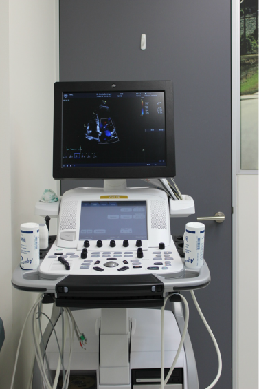
Definition
An echocardiogram is a test that uses sound waves to create a moving picture of the heart. The picture is much more detailed than a plain x-ray image and involves no radiation exposure.

Why the Test is Performed
This test is performed to evaluate the valves and chambers of the heart in a noninvasive way. The echocardiogram allows doctors to diagnose, evaluate, and monitor:
  • Heart murmurs
  • Abnormal heart valves
  • The pumping function of the heart for people with heart failure
  • Damage to the heart muscle in patients who have had heart attacks
  • Infection/inflammation in the sac around the heart (pericarditis)
  • Infection on or around the heart valves (infectious endocarditis)
  • The source of a blood clot or emboli after a stroke or TIA
  • Congenital heart disease
  • Atrial fibrillation
  • Pulmonary hypertension

How the Test is Conducted
A cardiologist or specially trained sonographer performs the test.
You will be asked to remove your clothes from the waist up and lie on an examination bed. Electrodes will be placed on your chest to allow for monitoring your heart rate and rhythm. An instrument called a transducer that transmits high-frequency sound waves is placed on your ribs near the breast bone. A transducer gel will also be used to obtain the images. This may be cold and is water based. It washes off easily at the end of the test.

Additional images will be taken at the left lower chest. The transducer picks up the echoes of the sound waves and transmits them as electrical impulses. The echocardiography machine converts these impulses into moving pictures of the heart. The Doppler probe records the motion of the blood through the heart.

An echocardiogram allows doctors to see the heart beating, and to see many of the structures of the heart.
The result will be discussed with you at the end of the test.

How to Prepare for the Test
There is no special preparation for the test.

​Risks
There are no known risks associated with this test.
echocardiogram.pdf
File Size: 77 kb
File Type: pdf
Download File

Picture
  • Home
  • About
  • Cardiologist Team
    • Dr Amely Zaininger
    • Dr Elizabeth Robertson
    • Professor Mark McGuire
    • Dr Caroline Medi
    • Dr Kevin Liou
    • Dr Kevan Karimi
    • Dr Lucy McGrath-Cadell
    • Dr Katy Ellenberger
    • Associate Professor Rebecca Kozor
  • Facilities and Services
  • For Patients
    • Stress testing
    • Echocardiogram
    • Exercise Stress Echocardiogram
    • Electrocardiogram
    • Ambulatory Blood Pressure Monitoring
  • For Doctors
  • Location and Contact